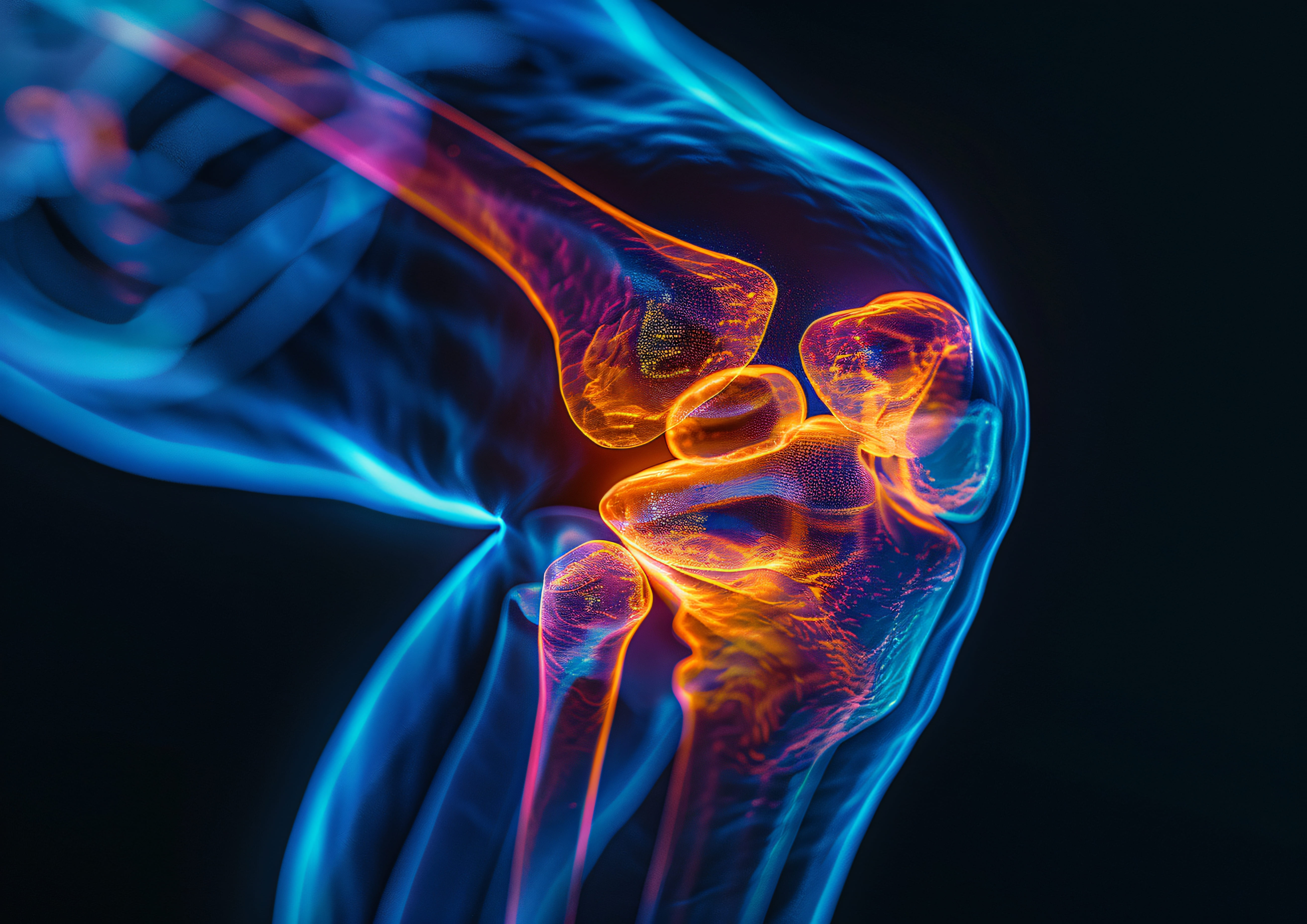
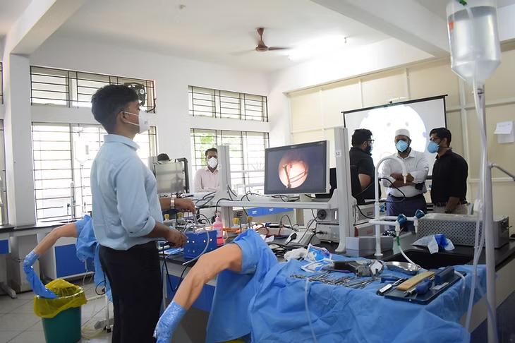
Hands-on Workshop on the Wet Knee Model & Cadaveric Shoulder
 2 May 2026
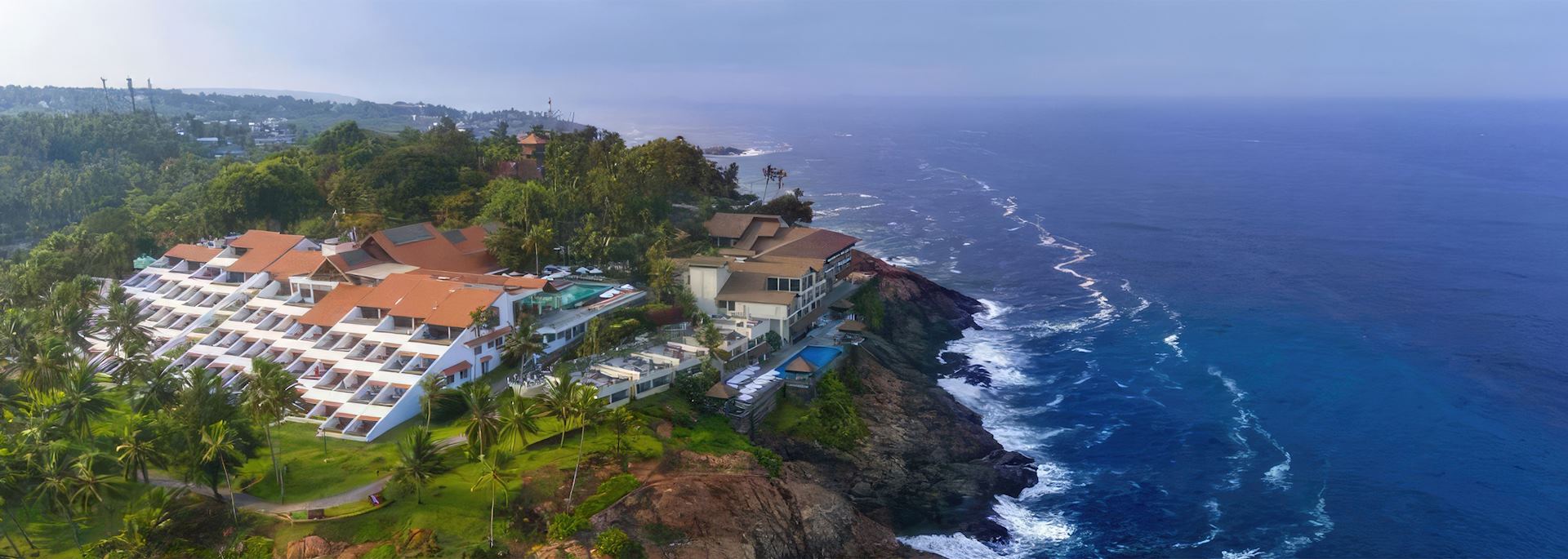
  Leela Hotel, Kovalam, Trivandrum
 Arthrolearn presents Trivandrum Arthroscopy course (TAC) 2026 on May 2nd and 3rd. Theme: Sports Surgeries


Introducing
Trivandrum Arthroscopy Course 2026
Theme : Sports Surgeries
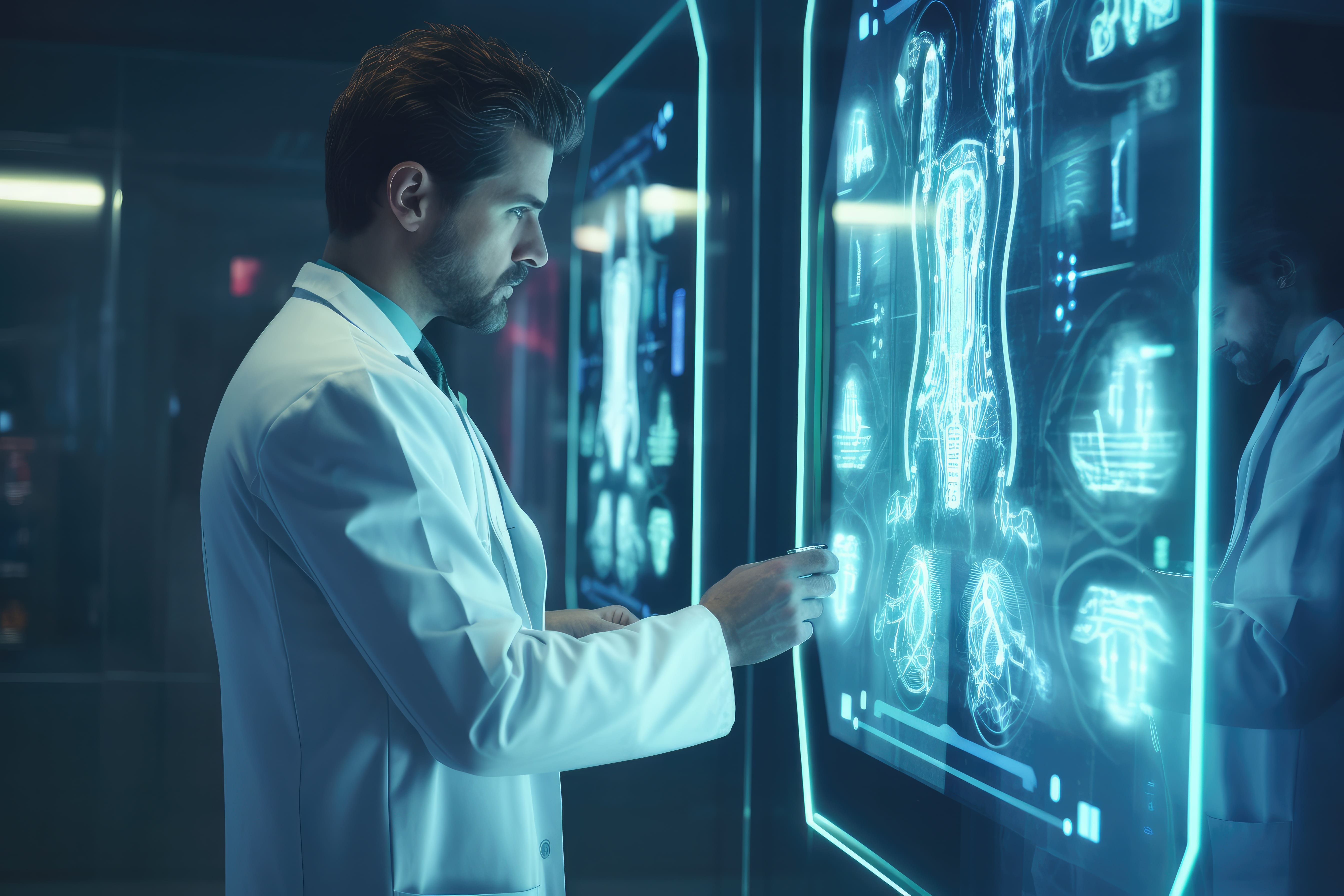
"Welcome to the future of Orthopaedics!"
Hands-on Workshop on the Wet Knee Model & Cadaveric Shoulder on May 2nd
days
hours
minutes
seconds
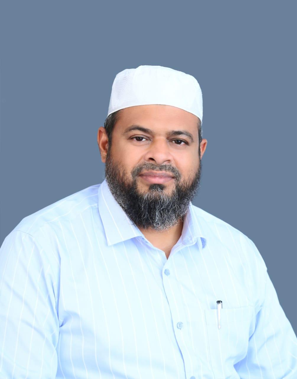
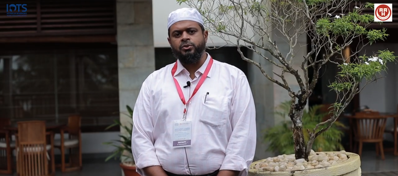
Dr. Shammas B.M
Course Chairman
(Al Arif Hospital TVPM)
Prof.Dr Romain Seil
Scientific Chair
(Luxembourg)
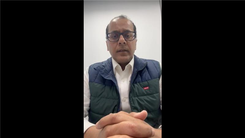
Prof.Dr Bashir Zikria
Course Co chair
(John Hopkins, USA)
Dr. Aizel Sherief
Course Coordinator
(Aspetar, Qatar)
Welcome Message
Dear Colleagues,
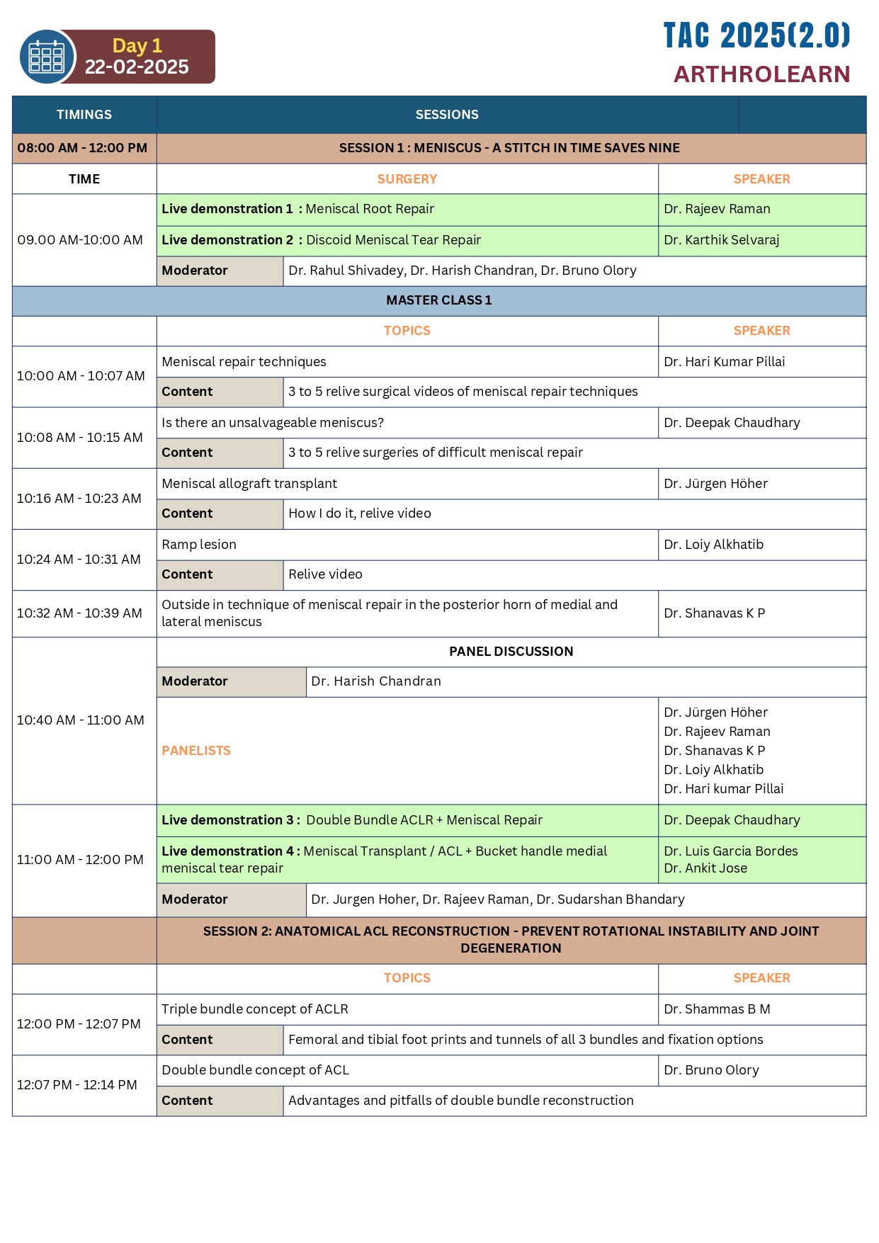
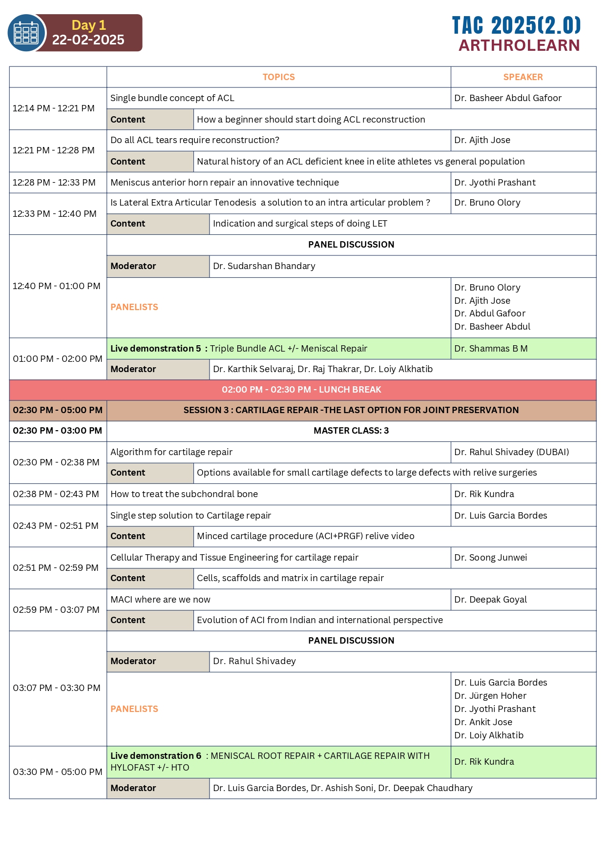
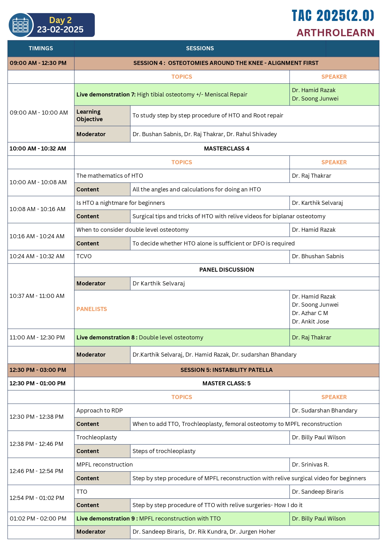
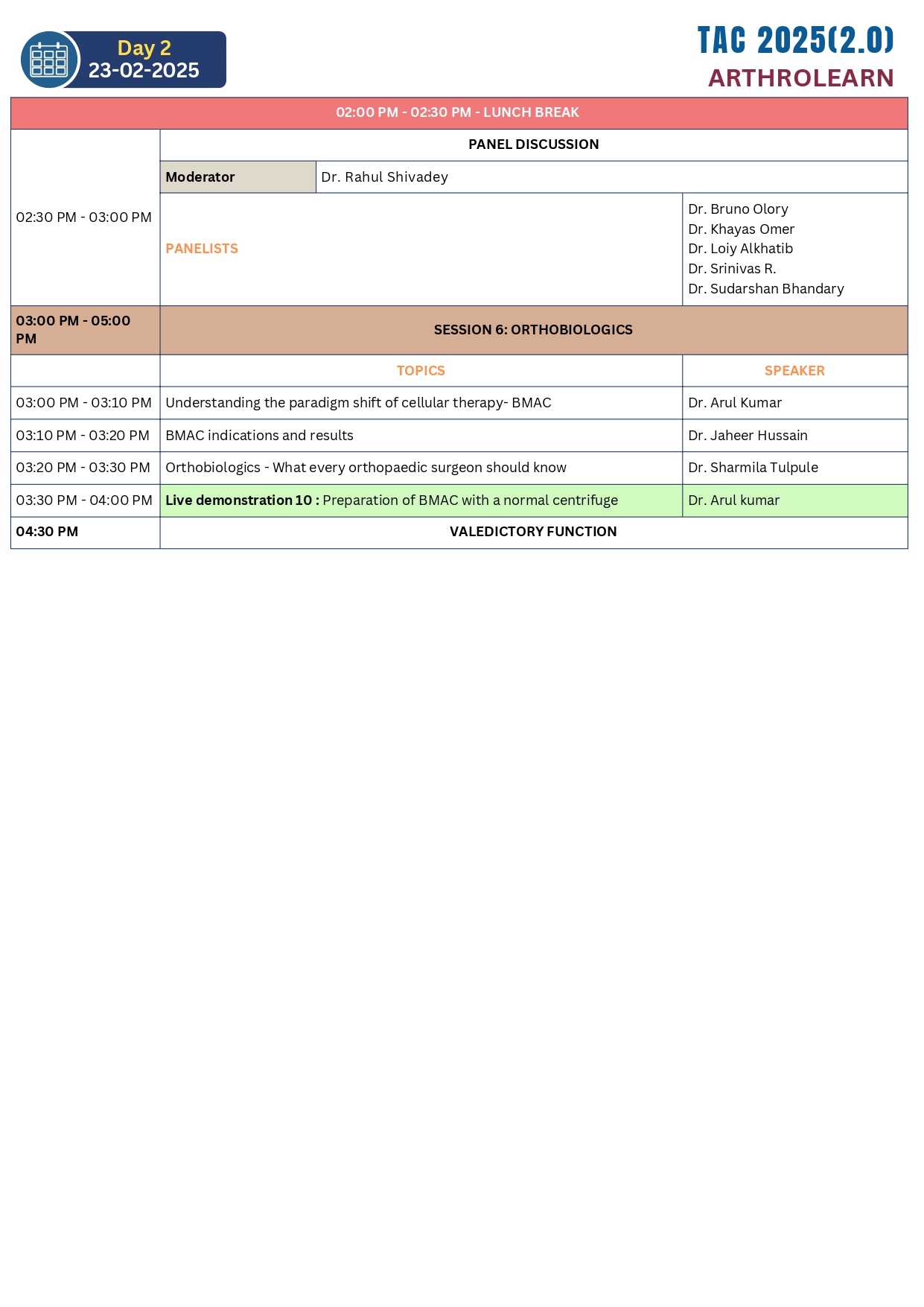
We are thrilled to announce the upcoming Trivandrum Arthroscopy Course, scheduled for May 2 & 3 2026, at the Leela Hotel in the beautiful coastal town of Kovalam. This serene destination, renowned for its stunning beaches and tranquil atmosphere, offers the perfect backdrop for an immersive learning experience. The course will provide an in-depth exploration of advanced arthroscopic techniques, featuring Twelve live surgeries that focus on innovative methods for Knee joint Preservation. Participants will engage in pre-operative planning sessions, where they will gain invaluable insights from leading experts in the field. Detailed discussions will complement the live demonstrations, allowing attendees to deepen their understan ding of the latest practices and techniques in arthroscopy.
This unique opportunity not only aims to enhance your skills but also to foster connections with peers and industry leaders, all while enjoying the breathtaking beauty of Kovalam. We look forward to welcoming you to this enriching event that promises to be both educational and inspiring.
Dr. Shammas B.M
Course Chairman
Speakers
Residential Packages
(for conference only)
The ArthroLearn Program
Since founding ArthroLearn in 2020, our mission has been to discover the world’s most innovative way to teach Arthroscopic Surgery.
Arthroscopic surgery is a developing area of Orthopaedics and Traumatology. In recent years, arthroscopy techniques including newer concepts of ACL/PCL reconstruction, Meniscal repair, articular cartilage, Meniscal Replacement surgery, orthobiologics, stem cells are being defined. Shoulder and ankle arthroscopy surgery continues to advance in leaps and bounds. We are advancing to a stage where even complex surgeries can be performed with least morbidity using an arthroscope and a dependable arthroscopy surgeon is essential in any Orthopaedic surgery team whether trauma or otherwise.
To take attention of our colleagues to this future trend and close the exposure deficit, and to encourage surgeons to do arthroscopy through a new scientific platform, we aimed at starting an Arthroscopy Training Initiative – “ARTHROLEARN” using customised simulator models where surgeons can learn to do various techniques in scopy before actually performing on a patient.
Our vision is to become a reputable Arthroscopy centre of high quality and a Training Centre to boost the surgeon’s confidence. We would like to thank you in advance for your scientific contribution towards this upcoming programme and appreciate it if you could give the largest support by participating in our initiative.
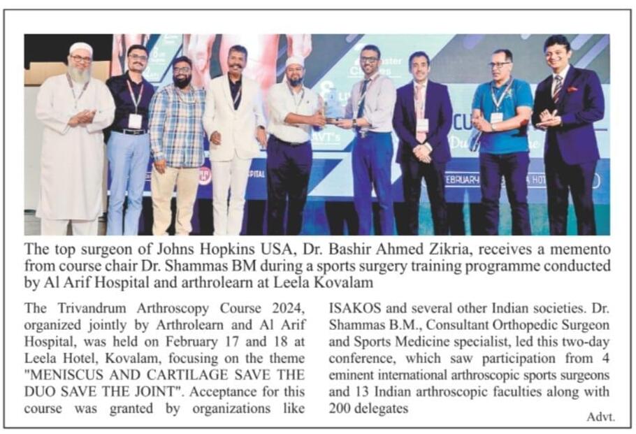
Making Headlines : Our Success Story
We are honored to have been featured in The Hindu newspaper for our efforts in advancing sports surgery training. The event, held at Leela Hotel, Kovalam, highlighted the theme: "Meniscus and Cartilage Save the Duo, Save the Joint."
This two-day conference brought together renowned experts, including international and Indian arthroscopic surgeons, to discuss advancements in arthroscopy techniques. It was a proud moment to showcase our dedication to innovation and excellence in the field of orthopedics.
The event saw the participation of 4 eminent international surgeons and 13 Indian faculty members, along with over 200 delegates.
As featured in The Hindu, February 2024.
ARTHROKNEE
ARTHROKNEE, an artificial wet knee simulator model was developed by Dr. Shammas B.M and Dr Vaishnav Satheesh in 2020, the patent application for which is pending.
The present invention provides a human anatomical knee model for arthroscopic procedures with critical anatomical structures important for a surgeon to identify and reference when performing knee arthroscopy and has the following components
Read MoreTestimonials
Dr Shammas B M
Dr Rahul Shivadey
Dr David Barastegui
Dr Ankit Jose
Dr Fayaz Memon
Dr Sundarajan S R
Dr Raju Eswaran
Dr Bashir Ahamed Zikria
Upcoming Workshop
2 May 2026 Leela Hotel, Kovalam, Trivandrum Nearby Places to Visit
Discover the charm of the locale with its rich heritage, inviting natural landscapes, and diverse culinary experiences. Immerse yourself in the cultural tapestry, explore nature's retreats, and savor local flavors, creating memories that linger long after your visit.
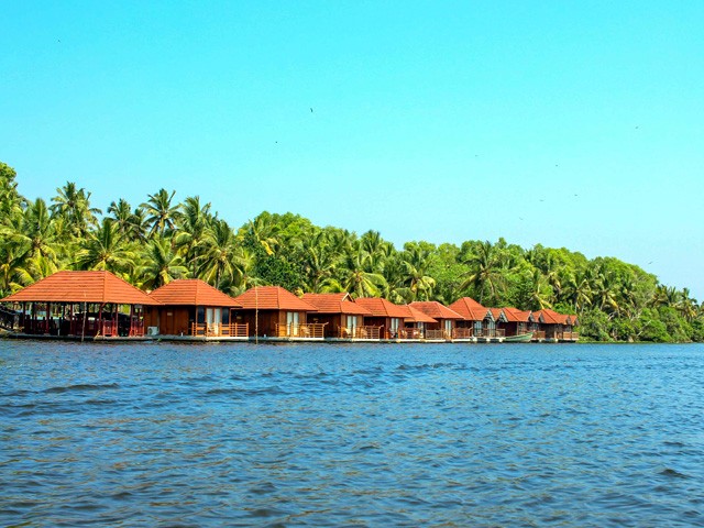
Poovar

Kovalam

Varkala
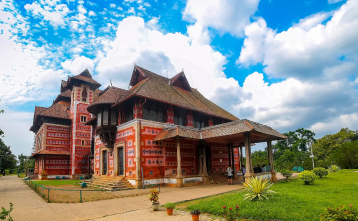
Napier Museum